Gynaecological examinations
Many women still fail to attend their gynaecological health checks and early detection screenings on a regular basis. These are so important because they can help detect possible gynaecological diseases in good time, allowing them to be treated.
Depending on the age of the woman, it is possible to have special health checks carried out. These examinations focus on, for example, identifying and treating cancer and its precursors before the cancer spreads and becomes life-threatening.
The main purpose of health checks is therefore to increase the chances of the patient being cured.
Health checks
A gynaecological examination includes a consultation on topics such as menstruation, hormone levels, sexual matters and contraception as well as a vaginal examination. It also serves to ensure the early detection of breast or cervical cancer. For example, a cervical swab is taken and the breast is check for lumps. Specific ultrasound examinations of the breast and abdomen may also be considered.
It is recommended that women attend these check-ups at the specified intervals. Aside from regular check-ups, these kinds of examinations are always also carried out when certain symptoms are already present and they need to be clarified in more detail. As a general rule, gynaecologist visits for the early detection of certain illnesses should be carried out at least once a year from the age of 20. As part of mammography screening programmes, all women over the age of 50 are also invited for a mammogram.
Ultimately, however, the frequency of visits to a gynaecologist depends on a woman’s individual risk of disease. For example, it is important for a patient to inform their gynaecologist if, for example, there is an increased incidence of cancer in their family. In this case, the treating doctor will discuss with the patient the intervals at which a gynaecological assessment would be advisable.
What symptoms should be checked for?
The gynaecological examination is primarily a preventive health check, which means that a woman undergoes screening even though her body does not show any signs of illness. However, if a woman is experiencing symptoms or has concerns, she should make a specific appointment with her gynaecologist.
Symptoms that are often the reason for a gynaecological examination include:
- Discharge from the vagina
- Period-related issues, e.g. pain during a period or a very severe and long period of menstrual bleeding
- Pain, burning or itching in the genital area, for example during sexual intercourse or urination
- Abnormal changes to the breast in the form of hardening or lumps
Causes
The causes of the symptoms listed above are manifold. Diseases that may cause them include:
- Endometriosis
- Fibroids in the uterus
- Premenstrual syndrome (PMS)
- Cystitis
- Breast cancer
- Cervical cancer
Consultation and urine sample
Before the actual physical examination begins, the patient is first asked to hand in a urine sample. The doctor then uses a special test strip to examine the urine for germs, blood or signs of inflammation. The presence of pregnancy can also be checked for by means of a urine test. In addition, an empty bladder makes the subsequent ultrasound examination easier.
In the next step and before the physical examination begins, the gynaecologist asks the patient about any possible symptoms and clarifies whether the patient’s immediate family has already had an increased incidence of breast cancer or cervical cancer in order to obtain possible indications relating to the family history. Additional discussions may include:
- The method of contraception currently used or the desire for contraception
- Their menstrual bleeding and their cycle and questions about the regularity, heaviness and duration of their period
- Possible occurrence of intermenstrual bleeding and/or vaginal discharge
- Information about their medication
- The presence of possible metabolic diseases
- Questions about sexual activity and partnership status
Physical examinations
Spotting external changes
The patient is asked to remove her clothing from the waist down and take a seat in the gynaecological examination chair.
- The gynaecologist takes a look at the woman’s external genital organs and, if necessary, to spot any external changes such as injuries, redness or abnormalities of the mucous membranes.
- The doctor also palpates the lymph nodes of the groin region and the patient’s abdomen.
Examination with medical instruments
- Speculum: For a closer examination of the vagina and the lower part of the cervix, an instrument called a speculum is used. A speculum is a medical instrument that typically widens body openings such as the vagina or anus so that the doctor can examine the inside of a woman’s vagina.
- Colposcopy: The doctor can also examine the vagina and the cervix during a colposcopy. A special microscope, called a colposcope, is used for this purpose. Colposcopy is mainly used for the early detection of cervical cancer and its precursors.
Bi-manual pelvic examination
- Bi-manual pelvic examination: Once the examination using these instruments has been completed, the bi-manual (with both hands) pelvic examination follows, whereby the gynaecologist first carefully inserts the index finger into the vagina to check the stretchability of the tissue and the presence of bulges, hardening or lumps. The doctor then places their second hand on the lower part of the patient’s abdomen from the outside, allowing them to feel the uterus. During this process, portio pressure tenderness is also checked. If the movement of the finger at the portio, the transition area between the vagina and the cervix, causes pain, this may indicate the presence of inflammation in the lesser pelvis.
- Examination of the rectum and posteiror wall of the uterus: In addition, the doctor inserts a gloved finger into the patient’s intestine in order to examine the rectum and the posterior wall of the uterus.
Pap test
As part of the vaginal examination, the gynaecologist performs a smear test known as the Pap test (also: cytological smear) for the early detection of cancer.
To do this, the doctor carefully removes individual, superficial cells from the cervix (the portio surface) using a spatula and a small brush from the cervical canal. The sample is then sent to a cytological laboratory. In the case of abnormal Pap findings, i.e. findings based on cell changes, precursors or even cancer cells, further investigations and examinations are necessary.
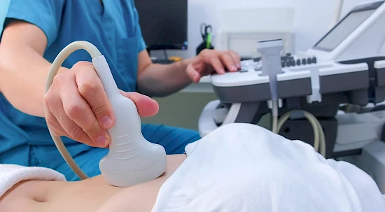
Ultrasound
At the end of the vaginal examination, an ultrasound examination is usually carried out using a special transducer which is coated in gel.
This is rod-shaped, allowing it to be inserted easily and painlessly into the vagina, giving the gynaecologist the opportunity to assess the wall and mucous membrane of the uterus, the cycle phase, the ovaries and the spaces of the lesser pelvis.
Breast examination and mammography
Palpation of the breasts
Once the patient has got dressed again, the gynaecologist will carry out a final breast examination. This is a palpation of the breasts with the aim of detecting any pain that may be present and feeling any lumps or hardening. As a rule, the doctor then shows the patient how she can regularly examine her breasts herself so that any changes can be detected at an early stage.
Mammography
From a certain age or if the gynaecologist deems a further investigation necessary, an X-ray examination of the breast, known as mammography, may also be ordered. This is a simple, quick X-ray examination of the female breast, which only takes a few minutes. The images (mammograms) can then be used to assess the normal, age-dependent condition of the breast and to distinguish this from pathological changes. In this case, tumours that cannot be felt with bare hands can be detected at an early stage.
Mammography is recommended every two years as part of a screening programme for early detection of breast cancer from the age of 50.
Procedure of the examination
- As part of a mammography, each breast is placed individually between two sheets of plexiglass, which are then briefly pressed together (known as compression) in order to spread out the breast for the recording. This unpleasant and sometimes very painful procedure is necessary to reduce the radiation dose and to obtain better images. The compression does not cause permanent damage to the breast.
- In principle, two images are taken of each breast, whereby the beam path runs once from top to bottom and once diagonally from the middle to the side.
Breast self-examination
After every visit to a gynaecologist, it’s a good idea to check your own breasts about once a month – just as your doctor has suggested.
Self-examination is easiest between the third and seventh days after the start of menstrual bleeding, as the mammary gland tissue is particularly soft at that time, which is why tissue changes are more noticeable.
FAQs
When is a gynaecological examination advisable for children?
An examination of young girls is necessary in the following cases, although an external examination of the genital organs is usually sufficient, which is why the vaginal palpation can be omitted:
- Bleeding from the vagina before the start of the menstrual period
- Pain, burning, discharge or itching in the genital area
- Suspicion of malformations
- Suspected developmental disorders
- Suspicion of sexual abuse
What happens when a smear is done?
The medical term “smear” refers to the removal of the body’s own material from the surface of wounds or mucous membranes by means of a sterile cotton swab, small brushes or small spatulas for microbiological and/or cytological diagnostics, i.e. the analysis of cell material for the detection of diseases or cell changes. The swab used as part of the gynaecological examination is called a Pap test.