

Open from Monday to Friday from 08:00 to 18:00 and on Saturday from 08:00 to 12:00
Address
Clinique Générale-Beaulieu
Chemin Beau-Soleil 20
1206 Geneva

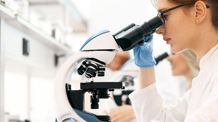
The radiology institute is ready to welcome you with a team of radiologists who offer high-quality services and skills in the most specialised imaging fields: neuroradiology, ENT radiology, female imaging, and thoracoabdominal, urologic, osteoarticular, vascular and interventional radiology.
Outpatient services are available to all patients, regardless of their insurance cover. They are subject to the same Tarmed tariffs as public institutions.
Radiologists are qualified to assess how useful the prescribed examinations will be in making the diagnosis your doctor is looking for. Indications are readily discussed with prescribing doctors in order to carry out the most suitable investigation for the situation, avoiding unnecessary examinations or too much radiation.
Those cases that require a multidisciplinary approach to define their treatment – senology (Centre du Sein – breast centre) and gynaecology, pulmonology, urology, oncology, and neurology (Neurocentre) – are discussed during set multidisciplinary meetings, which are organised at our institutes.
Cardiac MRI and CT angiography of the coronaries, the supra-aortic arteries, the aorta and its branches, including those of the lower limbs; venous Doppler ultrasound. Cardiac scintigraphy.
Full-body (MRI), lung (CT), brain (MRI, scintigraphy, PET/CT), densitometry, mammography, ultrasound scans, digital radiography.
Ultrasound, CT, MRI, study of swallowing, transits, enemas, study of the small intestine using enteroclysis, CT enterography, MR enterography, virtual colonoscopy using CT, standard defecography, MR defecography, gastric emptying and hepatobiliary scintigraphy, Octreoscan, PET/CT, biopsies, drainage of intra-abdominal collections. Insertion and care of percutaneous feeding tubes (PRG and PRJ), percutaneous intestinal stenting, drainage and stenting of bile ducts, removal of gallstones.
Neck ultrasound including thyroid and parathyroid glands; punctures; CT and MRI; thyroid and parathyroid scintigraphies; beta treatment of hyperthyroidism; Thyrogen tests to monitor thyroid cancers. Ablation of thyroid nodules using radiofrequency.
Drainage; spine and joint infiltrations, isotope treatment in the form of alpha therapy and beta therapy.
Abdominal and transvaginal ultrasound; pelvic and abdominal MRI; abdominal CT; bone densitometry with trabecular bone score.
Brain, spinal column, spinal cord and pituitary CT and MRI; CT angiography and magnetic resonance angiography of pre- and intracerebral vessels; infiltrations; cementoplasties. But also brain perfusion and specialised SPECT/CT, such as Datscan; 18FDG and fluorocholine PET/CT, and PET/CT with specific radiotracers for Alzheimer’s disease.
Standard radiography, ultrasound, CT scan, MRI. Performance of biopsies, cementoplasties, percutaneous thermal ablations (hepatic, renal, pulmonary, bone, etc.). Full-body and bone scintigraphies with specific radiotracers, and full-body PET/CT scans.
Study of swallowing; neck, facial bone and sinus CT scans; MRI of the neck and temporomandibular joints; CT dacryocystography; punctures; ultrasound; study of the salivary glands using ultrasound, CT and MRI; PET/CT; and scintigraphies. Minimally invasive treatment of head and neck pain.
Digital radiography of the musculoskeletal system; MRI; CT; MR and CT arthrography; ultrasound; infiltrations; biopsies; full-body and bone SPECT/CT.
Densitometry with trabecular bone score and vertebral morphometry, analysis of body composition, cementoplasties, vertebral augmentation.
Abdominal ultrasound including kidneys, bladder, pylorus and soft tissues; cranial transfontanellar ultrasound in infants; scrotal and hip ultrasound. Transits, enemas, voiding cystourethrogram, CT scan, MRI (depending on the child’s age), digital radiography including bone age, and all kinds of paediatric scintigraphies.
Digital radiography, pulmonary CT, biopsies, drainage. Bronchial embolisation and embolisation of pulmonary arteriovenous malformations. Pulmonary scintigraphy.
Mammography with tomosynthesis and double reading; screening mammography within the cantonal foundations’ framework; ultrasound; MRI; biopsies with ultrasound, stereotactic radiosurgery and MRI; lymphoscintigraphy.
Abdominal and endorectal ultrasound, CT and MRI, IVP (intravenous pyelogram), VCUG (voiding cystourethrogram), cystography, biopsy, and drainage. Insertion of percutaneous nephrostomies. Renal scintigraphy (MAG3 and DMSA), FDG and fluorocholine PET/CT, prostate MRI with navigation software for biopsies (Artemis).

Standard radiology or radiography is the study of joints at bone level. As a first-line examination, radiography makes it possible to visualise specific parts of the human body (bones and certain organs) and detects fractures and osteoarthritis. It uses X-rays (high-frequency waves), which – when passing through the body – are absorbed by different structures (e.g. bones, soft tissues). On X-rays, bones will appear white, while soft tissue will appear grey.

Interventional radiology combines both radiology (using X-rays) and disciplines such as surgery, medical oncology and urology. The advantage of this new technique is to carry out minimally invasive, imaging-guided procedures to:
a) Stop bleeding (by plugging the bleeding vessel from the inside)
b) Destroy tumours
Interventional radiology uses percutaneous techniques that, using different types of probes and medical imaging (scanners, MRI, ultrasound), locate and reach the tumour. Then, depending on the chosen technique and probe, the tumour will either be burned from the inside (heat emitted by radiofrequencies or microwaves) or frozen to -80 °C (cryotherapy: the liquid injected by the probe will freeze the cancer cells, which will eventually burst)
Even some previously incurable tumours can now be treated with a new technique called radioembolisation, where radioactive particles are injected into the tumour and irradiated from the inside until it is destroyed.
c) Another very promising technique is to inject modified viruses into the tumour, which will then only attack the cancer cells. Naturally, the virus is modified in such a way that it cannot develop into healthy cells.

The CT scanneris a device that rotates around the patient using an X-ray tube. Sensors scan all or part of the human body and provide data to reconstruct the image digitally. CT scans can also be used for biopsies, drainage and radiofrequency cancer treatments (interventional radiology: destruction of the tumour using heating or cooling).
For irradiation, only low doses are used and every precaution is taken to limit the exposure to X-rays in the area under examination to an absolute minimum. For example, a single image corresponds to the average natural (cosmic) exposure during a four-hour flight. However, for pregnant women, special precautions must be taken on a routine basis. This is why it is important to notify the medical team about any potential pregnancy.

Mammograms, recommended for all women between the ages of 50 and 75 years, are designed to detect abnormalities in breast tissue as early as possible, before they even cause clinical symptoms. This allows breast cancer to be detected, ruled out or diagnosed before it becomes palpable. This radiological examination has evolved considerably in recent years, therefore the doses given during the examination are extremely low. Mammograms are equipped with tomosynthesis, a technique allowing for sectional imaging, which improves the resolution of the image produced and facilitates the detection of any abnormality.
During the mammogram, which only lasts a few minutes, the patient will be accompanied by an experienced radiologist who will explain the procedure in detail and answer any questions she may have. For a so-called diagnostic examination, it is likely that the patient will undergo an ultrasound scan in addition to the mammogram.
The patient will receive the results of the mammogram by post within eight working days. The results will also be sent to the GP whose name was given on the health questionnaire. This is sufficient time for two experienced radiologists to analyse and interpret the mammogram in detail, so that no abnormalities are missed.
Most of the Swiss Medical Network’s radiology centres actively support and collaborate in breast cancer screening. These examinations are coordinated by screening centres or cantonal bodies (Swiss Cancer Screening).

Bone densitometry (or osteodensitometry or mineralometry) is a radiological technique that measures bone mineral density to determine bone strength. It thus makes it possible to:
Sites measured are typically the lumbar spine, proximal femurs (hips and femoral necks) and sometimes forearms (radius). In rare cases, a full body measurement may also be taken. During the examination, using the same apparatus, lateral spine imaging (vertebral morphometry) is usually carried out in order to look for any spinal defects or compression fractures. Furthermore, it is important to be able to assess not just the amount of calcium in the bones (quantitative) but also its distribution (qualitative) by analysing vertebral microarchitecture.
This is done using a software programme that allows for a vertebral trabecular bone score to be calculated. Based on all these parameters and a targeted medical questionnaire, the 10-year fracture risk may be determined for all major osteoporotic fractures or hip fractures. These are essential factors in establishing when and which treatment should be put in place.

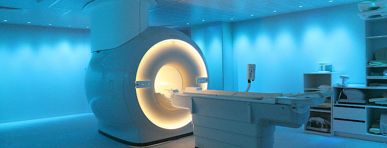
Magnetic resonance imaging (MRI) is a technique that does not use X-rays. The device consists of a magnet characterised by its power (1, 1.5 or 3 Tesla) and an examination bed which glides down its centre. The examination creates images of the human body thanks to the hydrogen atoms it contains. In a strong magnetic field, all hydrogen atoms move towards this field. They are then excited by radiofrequency waves for a very short period of time. When the wave emission stops, the atoms return to the equilibrium state and the accumulated energy produces a signal that is recorded and processed as an image. Specific antennas according to the area studied are connected and used to collect the emitted signal. These signals are mathematically analysed to produce images.
According to the technique or sequences, MRI enables the morphology of the organs studied to be brought to light in a detailed way. It demonstrates an increased sensitivity in detecting different pathologies of the soft tissues, making vessels, cartilage and nerve pathways stand out. The acquisition of three-dimensional images allows for their reconstruction on all planes.

This painless and non-radiation imaging technique allows a diagnosis to be made or a rapid intervention to be carried out alongside the patient. It can be repeated as often as necessary without any health risks. It is very useful in many fields of radiology (ENT, breasts, musculoskeletal system, blood vessels, liver, gall bladder, pancreas, kidneys, bladder and pelvis).
Technically, it is a beam of ultrasound whose frequency is adapted to the organ to be examined. This beam is reflected more or less depending on the anatomical structures it encounters. In turn, this allows for an image of the organ in question to be reconstructed. It is shown directly on the screen of the apparatus for an immediate and dynamic interpretation.

A PET/CT scan is used for oncological, cardiological and neurological examinations and to investigate sources of infection. In oncology, this technique can be used to confirm the presence of a cancer. As the patient’s whole body is examined, it can also detect tumour cells present in other organs (metastasis). With the PET/CT scan, quantitative values can be obtained and used to track tumour cells in order to determine if a treatment is working and evaluate if the cancer is improving.
From a therapeutic point of view, nuclear medicine uses radioactive pharmaceuticals to destroy diseased cells. A pre-treatment scintigraphy or PET/CT scan ensures the radiation correctly targets the diseased cells. Afterwards, the patient is administered radioactive pharmaceuticals that are specific to the cells that need to be destroyed.

The device records the gamma rays emitted by the patient to create functional images. Activity curves can be generated, for example to map kidney function or compare the activity of a body part on the left with a body part on the right. This is often used to examine stress fractures or bone disorders.
Two radioactive tracers are used to examine the lungs; one is inhaled and the other injected. This technique is used to explore lung function before an operation, in cooperation with a pulmonologist, and to detect pulmonary embolisms.
To confirm a diagnosis of Parkinson’s disease, a cerebral scintigraphy scan may be carried out at the request of a neurologist.
In endocrinology, scintigraphy can be carried out on the thyroid or parathyroid glands. This is the preferred examination for hormonal issues, or to investigate thyroid or parathyroid nodules.
In the case of symptoms that can suggest coronary artery disease (feelings of suffocation, chest pain, shortness of breath), cardiac scintigraphy is indicated. For this examination, a cardiac stress test is carried out with a cardiologist once the radiotracer is administered. The result shows the activity of the cardiac muscle and allows doctors to diagnose ischaemia or infarct.
Open from Monday to Friday from 08:00 to 18:00 and on Saturday from 08:00 to 12:00
Clinique Générale-Beaulieu
Chemin Beau-Soleil 20
1206 Geneva


