
With a clear view ahead, you can overcome any challenge - take care of your vision!
Book your free laser consultation today and discover how you can start a new chapter in your life without visual aids.
Even in early childhood, doctors examine your eyes. This is done routinely by the paediatrician who, will refer the child to an ophthalmologist if they identify any problems. In adolescence, it is often the teenagers themselves who complain of visual issues and are then examined in more detail by an ophthalmologist.
In adulthood, people go to an ophthalmologist regularly because age-related visual impairments or eye diseases can develop, for example due to excessive intraocular pressure. How often you should consult a specialist depends, of course, on whether you have problems with your eyes or if you suffer from certain illnesses.
If you have no eye problems, you should still have them checked at regular intervals.
An ophthalmologist should always be consulted immediately in the following situations:
Important: As a general rule, it is not possible for someone who is not a medical professional to distinguish between harmless changes in the eye and serious, potentially dangerous eye problems, which is why it is strongly recommended to consult an eye specialist in the event of any changes to the eyes in order to exclude or prevent long-term damage to the eye(s).
Ophthalmological emergencies include injuries and acute illnesses that may lead to permanent damage and impaired vision if they are not treated immediately.
If vision problems occur suddenly, this is usually an emergency that requires immediate treatment by a specialist. And it goes without saying that sudden blindness is always an absolute emergency.
The term “acute onset visual impairment” refers to all pathological changes in optical perception. These include sudden diminished visual acuity, impaired visual fields, flickering in front of the eyes, flashes of lightning and seeing double. The various causes of visual impairment range from eye diseases and neurological disorders to tumours.
If you suddenly see light streaks or small dark spots that float away when your eyes move, you should have someone take a closer look. The “mouches volantes” (from the French meaning “flying flies”) often turn out to be vitreous opacity, which is usually not dangerous. Larger dark spots and flashes of light in your vision, on the other hand, are an alarm sign that must not be ignored under any circumstances. The vitreous humour may have detached itself from the retina, resulting in a tear in the retina. Or eye inflammation (known as uveitis) is also responsible for these symptoms.
In rare cases, symptoms like these may also indicate retinal detachment. If a veil or “wall” appears in front of the eye (with or without “mouches volantes”, but in any case along with visual impairment), this may be the first sign of retinal detachment.
Dry eye is unpleasant and can have many different causes. They are often associated with ageing, wearing contact lenses or allergies. If left untreated, they can lead to chronic inflammation. For this reason, it is advisable to consult an ophthalmologist if you suspect that your eyes are dry (known as sicca syndrome).
Glaucoma is a silent, degenerative disease of the optic nerve, representing the leading cause of irreversible blindness in the West. It is generally characterised by high intraocular pressure that crushes the optic nerve, gradually destroying the nerve fibres that connect the retina to the brain. The reasons for the increased eye pressure are still unknown.
If not detected in time, glaucoma leads to a progressive and insidious reduction in the visual field, often noticed by the patient very late in life, and ultimately to loss of sight.
Cataract disease is characterised by an opacification of the lens, which is normally clear and transparent. The crystalline lens is a natural lens located behind the iris, which allows light rays to focus on the retina. When the crystalline lens loses its transparency, light can no longer penetrate to the back of the eye, resulting in a progressive loss of vision and sharpness of contours.
If left untreated, no improvement is possible and the cataract can lead to a slow deterioration of vision. With surgical treatment, visual improvement can be expected in the absence of other eye diseases.
The retina plays an important role in vision by transmitting light pulses from the eye to the brain via the optic nerve. The macula is part of the retina and is responsible for detailed vision. It is therefore important to protect the retina and the macula and to treat them effectively in the event of a retinal or macular disease.
Diseases of the cornea come in many variations and can affect all the different layers of the cornea. Corneal diseases are often accompanied by the sensation of having a foreign body in the eye or increased glare, whereby the loss of transparency of the cornea can even lead to loss of vision. A wide variety of forms of inflammation, eye injuries or age-related and congenital diseases can be responsible for this.
The retina and macula are the most sensitive parts of the eye and the most threatened by diabetes. The excess of sugar in the blood, caused by diabetes, weakens and damages the vessel walls of the retina. This failure of the vascular network prevents a good irrigation of the retina and leads to the death of nerve cells. In addition, the capillaries become more permeable and cause haemorrhages (non-proliferative diabetic retinopathy) or oedema (diabetic macular oedema). In advanced stages, new abnormal and fragile vessels may appear on the retina and further increase the risk of bleeding into the eyeball (proliferative diabetic retinopathy).
Laser treatment of diabetic retinopathy has shown excellent results and helps prevent the complications associated with this disease. Intravitreal injections are successfully used in the treatment of diabetic macular edema. Optimal control of diabetes and other risk factors, such as blood pressure and circulating fat levels (lipids), are also important in maintaining a healthy fundus.
Diabetes can also affect other parts of the eye. For example, it can cause cataracts to develop more quickly because of high levels of sugar in the eye. The blood vessels that supply the eye muscles can also be damaged by diabetes.
Diabetes damage in the eye often remains silent for a long time and does not cause any pain. It can be very severe without any visual impairment, which is why it is important to be checked by an ophthalmologist as soon as the diagnosis is made, followed by regular check-ups as recommended by the specialist.
The vitreous humour is a transparent jelly substance that fills the cavity inside the eye. As pathological changes to the vitreous humour are usually closely related to a retinal disease, it is recommended that any changes to the vitreous humour be examined by an ophthalmologist.
Eye inflammations are common and can be caused by a variety of factors. The eye is constantly exposed to pathogens and environmental stimuli. Inflammation develops when the eye is no longer able to defend itself against these kinds of harmful aggressors.
Normally, both eyes look in the same direction, but get a slightly different picture. These different images provided by the eyes are compared and assembled by the brain, resulting in a single, uniform image. When a person has strabismus, on the other hand, the eyes do not look in the same direction. They have a different resting position, which is why the brain cannot properly merge the images.
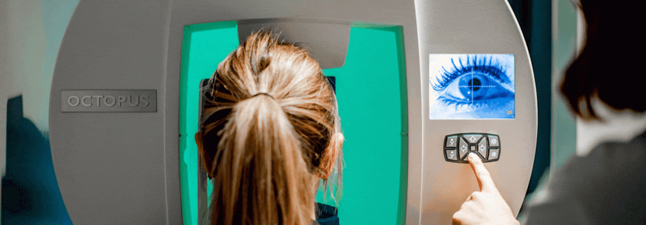
An ophthalmologist’s examination usually follows a specific structure: After talking about the nature and severity of the patient’s problems, an eye test is often carried out to check the visual acuity of each eye. The doctor then examines the eyes and takes a closer look at them using a slit lamp (this is a microscope that emits a narrow, slit-shaped light used for examinations). If necessary, the intraocular pressure is measured with the slit lamp. For some tests, eye drops are administered to dilate the pupil for a certain period of time. This makes it easier to assess the back of the eye and, in particular, the condition of the optic nerve.
A general eye check-up also includes a test of pupil reactivity. The pupil normally contracts reflexively when exposed to light. Additional tests relate to colour vision, where the patient is asked to recognise certain patterns on colour charts, eye motility and the field of vision. The ophthalmologist also looks at the eye and checks whether the conjunctiva or the eyelids are red or inflamed.