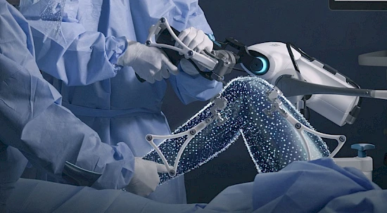
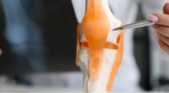
Hip prosthesis

The use of a hip prosthesis is the most successful treatment for painful osteoarthritis of the hip. The use of an artificial hip joint is considered when conservative treatment is no longer possible.

Hip prosthesis operations are now routine interventions and enable the function of the hip joint to be restored.

Symptoms

Hip problems are highly dependent on age and daily activities. They coincide with certain movements and often go hand-in-hand with restricted mobility.

In older people, hip pain usually occurs in connection with osteoarthritis, (deterioration of the cartilage) of the hip joint.

Causes

When the hip joint is so severely damaged or worn that conservative treatment can no longer be carried out, hip prosthesis is always used. Severe wear and tear of the hip joint results from degenerative changes, diseases or injuries to the joint.

Treatments

With surgical treatment, an operation is carried out to remove the diseased femoral head and parts of the femoral neck.

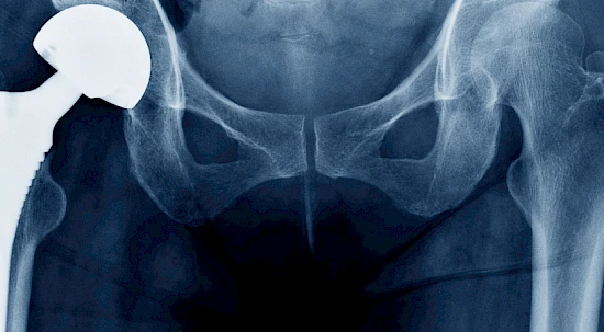
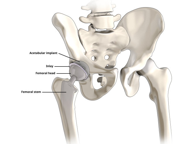
The natural hip socket is then prepared so that a new artificial hip socket can be anchored to it. The prosthetic head is then attached to an artificial shaft and the artificial hip joint is constructed.

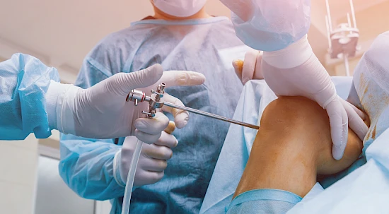
Thanks to the advanced method of keyhole surgery, which helps to preserve the tissue, the stabilising hip muscles are only pushed to the side, rather than being severed as they were before. In this minimally invasive procedure, the artificial hip joint is introduced from the front. An incision of about eight centimetres is made in the skin, through which the artificial hip joint is inserted without injuring the tissue.

The hip operation takes up to two hours and the hospital stay is around five days.

What happens during a hip prosthesis operation?

  1. Exposure of the hip joint: Minimally invasive procedure in which the affected hip joint is exposed without damaging the tissue, and the joint capsule is removed.
  2. Removal of the femoral head: A saw is used to separate the worn femoral head and femoral neck from the femur itself.
  3. Preparation and replacement of the hip socket: The surgeon begins reconstructing the hip using the new artificial hip joint. The hip socket is prepared and the old capsule and cartilage tissue are removed from the socket. Then the new hip socket and the corresponding inlay (cartilage replacement) are inserted.
  4. Preparation of the femur and insertion of the shaft: The hollow interior of the femur (marrow cavity) is prepared. Trial hip repositioning – the stability of the hip is tested using trial components. If stability is guaranteed, the new hip prosthesis is used. If the quality of the bone means that the it cannot be anchored without cement, then the prosthesis is cemented in place.
  5. Repositioning of the new artificial hip: The artificial hip joint is repositioned and the flexible connection between the shaft and socket is restored.

Aftercare

Once the operation is finished, the patient is required to stay in hospital for another five days.

On the first day after the operation, physiotherapy begins with gait training. In the first six weeks, crutches must be used to take pressure off the hips and protect the soft tissue.

After leaving the clinic, physiotherapy takes place over several weeks, which involves movement training and exercises to help rebuild muscle.

Services for patients living outside Switzerland

GENOLIER PATIENT SERVICES is at your side if you're looking to find the top specialists for hip prostheses in Switzerland.

Find out more about our specific offer for international patients and request your personalised programme today.

Our team is proud to offer services in multiple languages to meet the diverse need of our international clientele.

Français | English | Deutsch | Русский | Italiano | 中文

FAQs

What are the advantages of this soft tissue-focussed method?

Because, unlike with other methods, this technique does not injure the muscles, the patient experiences significantly less pain after the operation. As the procedure protects the tissue, blood loss is also relatively low. That also means that there is absolutely no need for the autologous blood donation, which was required for previous methods.

How is a hip constructed?

The artificial hip joint consists of four components. Two of these are directly connected to the bone: the acetabulum is connected to the pelvis and the shaft fits inside the femur. The new femoral head and the lining of the acetabulum, also called the inlay, form the new cartilage.

How long does an artificial hip joint last?

Although we might want them to last a lifetime, it’s important to be aware that an artificial hip joint is only ever a temporary solution. This can be caused by wear and tear on the sliding pair, especially if plastic or metal is used, or by the natural progression of the ageing process (osteoporosis), which can have a significant impact on the stability of the artificial joint.

Until now, artificial hip joints were expected to have an average lifespan of at least 10 to 15 years. Thanks to the tissue-sparing surgical method and the materials available today (e.g. ceramic-ceramic sliding pairs), we are confident that the lifespan of an artificial hip joint is now significantly longer. Of course, this is assuming that infections, fall-induced fractures or rapid progression of osteoporosis do not cause premature degeneration of the artificial hip joint.

What movements are allowed after hip surgery?

  • Hyperextension of the operated hip
  • External rotation of the operated leg
  • Crossing of the operated leg
  • Rotation of the upper body towards the healthy side

We find a specialist for you