-
Hospitals
back
Hospitals
- Clinique de Genolier
- Clinique de Montchoisi - Lausanne
- Clinique de Valère - Sion
- Clinique Générale Ste-Anne - Fribourg
- Clinique Générale-Beaulieu - Geneva
- Clinique Montbrillant - Chaux-de-Fonds
- Clinique Valmont - Glion sur Montreux
- Hôpital de La Providence - Neuchâtel
- Hôpital de Moutier
- Hôpital de Saint-Imier
French-speaking Switzerland -
Centres
back
Centres
- Centre Médical Eaux-Vives
- Centre Médical Valère
- Consultations dans le Haut Valais
- Médicentre Corgémont
- Medicentre Courroux
- Medicentre Courtelary
- Médicentre Moutier
- Médicentre Tavannes
- Medicentre Valbirse
- Montchoisi Medical Center
- Policlinique de Valère
- Polyclinic Genolier
- Ärztezentrum Bümpliz
- Ärztezentrum Ittigen
- Ärztezentrum Oerlikon
- Ärztezentrum Ostermundigen
- Ärztezentrum Schönburg
- Ärztezentrum Siloah Liebefeld
- Ärztezentrum Siloah Murten
- Ärztezentrum Solothurn
- Ladies Permanence Stadelhofen
- Medizinisches Zentrum Biel
- Medizinisches Zentrum Haus zur Pyramide
- Xundheitszentrum
French-speaking SwitzerlandGerman-speaking Switzerland - Swiss Visio
- Medical specialty
- Doctors
-
For patients
- About us
-
Integrated care
-
Hospitals
back
Hospitals
- Clinica Ars Medica - Gravesano
- Clinica Sant'Anna - Sorengo
- Clinique de Genolier
- Clinique de Montchoisi - Lausanne
- Clinique de Valère - Sion
- Clinique Générale Ste-Anne - Fribourg
- Clinique Générale-Beaulieu - Geneva
- Clinique Montbrillant - Chaux-de-Fonds
- Clinique Valmont - Glion sur Montreux
- Hôpital de La Providence - Neuchâtel
- Hôpital de Moutier
- Hôpital de Saint-Imier
- International Patients
- Privatklinik Belair
- Privatklinik Bethanien - Zürich
- Privatklinik Lindberg - Winterthur
- Privatklinik Obach - Solothurn
- Privatklinik Siloah
- Privatklinik Villa im Park, Rothrist
- Rosenklinik Rapperswil
- Schmerzklinik Basel
- Spital Zofingen
-
Centres
back
Centres
- Ars Medica Agno
- Ars Medica Bellinzona
- Ars Medica Manno
- Ärztezentrum Bümpliz
- Ärztezentrum Ittigen
- Ärztezentrum Oerlikon
- Ärztezentrum Ostermundigen
- Ärztezentrum Schönburg
- Ärztezentrum Siloah Liebefeld
- Ärztezentrum Siloah Murten
- Ärztezentrum Solothurn
- Centre Médical Eaux-Vives
- Centre Médical Valère
- Centromedico
- Consultations dans le Haut Valais
- Ladies Permanence Stadelhofen
- Médicentre Corgémont
- Medicentre Courroux
- Medicentre Courtelary
- Médicentre Moutier
- Médicentre Tavannes
- Medicentre Valbirse
- Medizinisches Zentrum Biel
- Medizinisches Zentrum Haus zur Pyramide
- Montchoisi Medical Center
- Poliambulatorio Pediatrico
- Poliambulatorio Sant'Anna
- Policlinique de Valère
- Polyclinic Genolier
- Xundheitszentrum
- Cancer centres
- Radiology centres
- Spine centres
- Sports medicine centres
- Neurology centres
- Centre du Sein
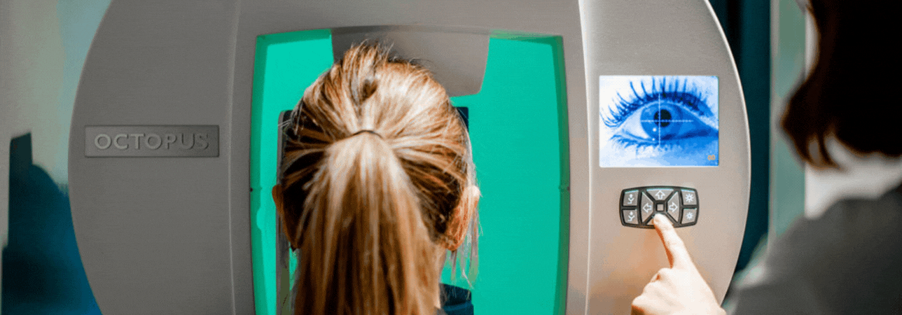
- Swiss Visio
- Medical specialty
- Doctors
-
For patients
back
For patients
- About us
-
Integrated care
close search